Regeneration solution provider
Global biomaterials manufacture
S1
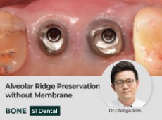
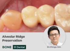
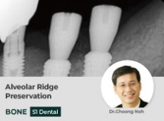
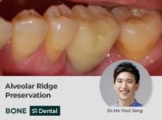
Alveolar Ridge Pres…
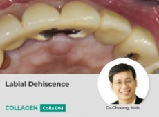
Despite of single use of S1 without membrane covering for ARP, Extra-fine soft tissue healing and full coverage was foun…
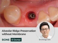
Alveolar Ridge Pres…
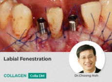
Not only volumatic change was found in CT scan, but also clinical situation for implant placement was acceptable. Additi…
Alveolar Ridge Pres…
For 5 months, Soft tissue and bony contour was well preserved in CT scan. But regenerated bone shows soft bone quality d…
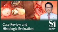
Alveolar Ridge Pres…
At 6 months after S1 bone graft material use, the boundary line between the S1 bone graft material and the natural alveo…
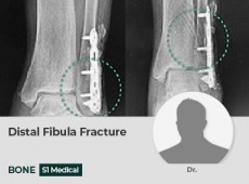
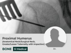
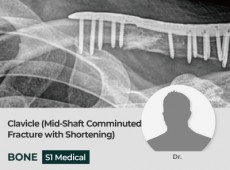
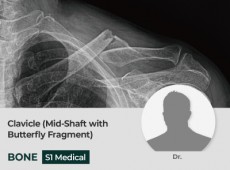
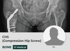
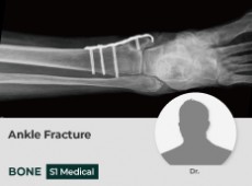
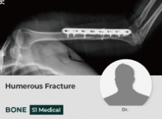
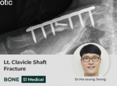
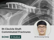
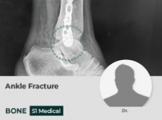
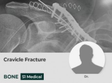
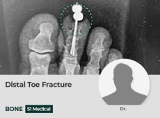
Distal Fibula Fract…
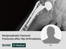
S1 Medical bone graft after fixing plate and screw with fracture caused by injury during exercise
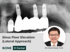
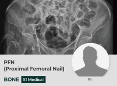
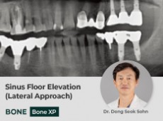
Sinus Floor Elevati…
A case that shows the formativeness of a space by lifting the sinus floor membrane using S1 bone graft material
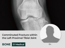
HTO (High Tibial Os…
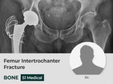
S1 Medical bone graft after HTO surgery to correct bent leg due to degenerative arthritis
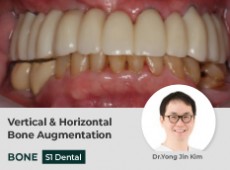
Vertical & Horizont…
Using a guided bone regeneration technique that has been widely performed, the biopsy shows good ossification and good b…
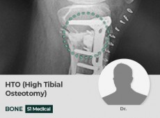
HTO (High Tibial Os…
S1 Medical bone graft after HTO surgery due to varus deformity due to degenerative arthritis.
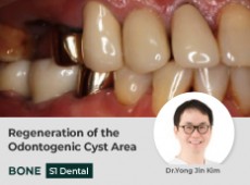
Regeneration of the…
After 3 months of using S1 bone graft material, a dense density of new bone was observed on radiographs.
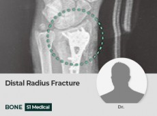
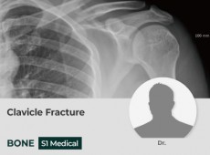
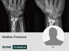
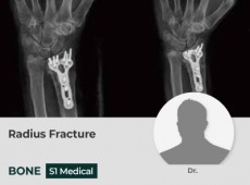
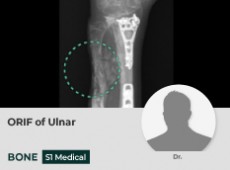
Distal Radius Fract…
S1 Medical bone graft after fixing plate and screw after complaining of wrist pain due to Distal Radius fracture from…
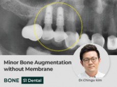
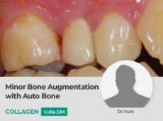
Minor Bone Augmenta…
Abundant bone regeneration was found after 3 Months, even regenerated bone covered over the cover screws. Regenerated bu…
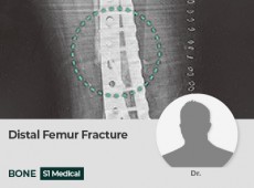
Distal Femur Fractu…
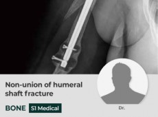
S1 Medical bone graft after fixing plate and screw from complaining pain due to Distal Femur fracture by accident
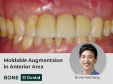
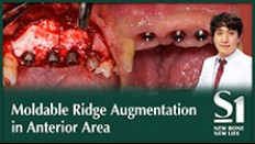
Moldable Augmentaio…
Observation of good healing of the soft tissue around bone graft material although the surgical area was wide.
Alveolar Ridge Pres…
Vertically augmented alveolar bone around the #36 implant can be confirmed on comparing the preoperative X-ray and posto…
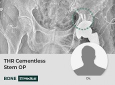
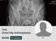
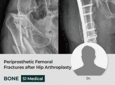
THR Cementless Stem…
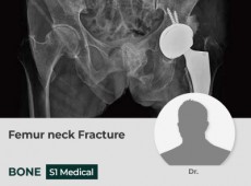
S1 Medical bone graft after total hip replacement surgery due to Acetablar defects
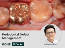
Periodontal Defect …
Although the membrane was not used, the volume of the bone graft material was maintained well and the new bone was well…
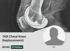
TKR (Total Knee Rep…
S1 Medical bone graft after total Knee replacement surgery due to Dista Femur Axis hole
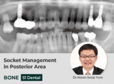
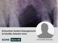
Socket Management i…
Even though the defect area was large, the shape was well maintained without a membrane, and new bone was formed well.
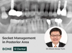
Socket Management i…
Even though the defect area was large, the shape was well maintained without a membrane, and new bone was formed well.
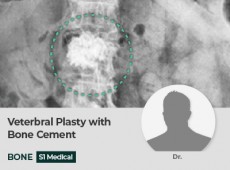
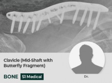
Veterbral Plasty wi…
Used with S1 Medical and bone cement for VP(Veterbral Plasty) surgery due to Verterbral fracture
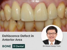
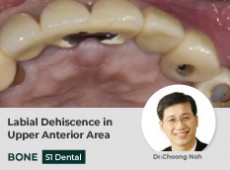
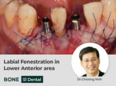
Dehiscence Defect i…
Though the bone width of the defect part was insufficient and the range was wide, the desired shape was well made after …
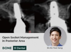
Open Socket Managem…
Though it was sutured in an open wound state immediately after implant placement with bone graft, secondary healing occu…
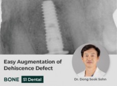
Easy Augmentation o…
Despite the lack of bone width in the defect area and in the anatomical structure that made it difficult to maintain the…
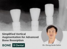
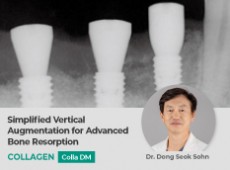
Simplified Vertical…
Though there is a wide range of bone loss at defect area, it can stably apply S1 bone graft material to the desired reg…
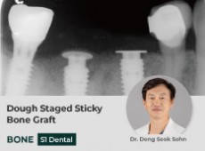
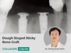
Dough Staged Sticky…
On radiographs 3 months after surgery, it was observed that the shape of the marginal bone was harmoniously connected wi…
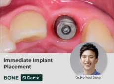
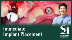
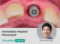
Immediate Implant P…
Used S1 and COLLA, the preservation of periodontal tissue was better than before extraction, and esthetically satisfacto…
BONE REGENERATION
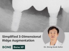
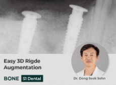
Simplified 3-Dimens…
After 3 months of surgery, it was confirmed by radiograph that the horizontal bone augmentation was done well
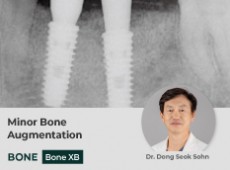
Minor Bone Augmenta…
After 3 months of using BOSS, bone formation and soft tissue healing without inflammatory response were observed
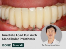
Imediate Load Full…
Cortical bone density was high after a long time after tooth extraction, but new bone was well formed
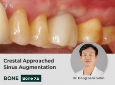
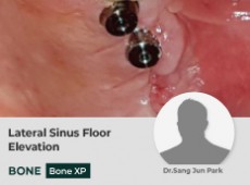
Crestal Approached …
Using a vertical approach, the base of the maxillary sinus was lifted and only a part of the fixture was fixed to the re…
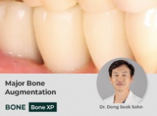
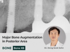
Major Bone Augmenta…
Cortical bone density was high after a long time after tooth extraction, but new bone was well formed
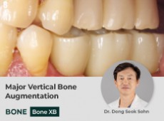
Major Vertical Bone…
The BOSS bone graft material was stably spaced despite the extensively performed vertical and horizontal bone augmentati…
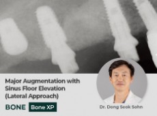
Major Augmentation …
Even though the inflammatory implant was removed, it was visually observed that the new bone formation was successful in…
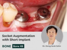
Socket Augmentation…
Considering the distance from the mandibular nerve to the mandibular posterior molar, the implant surface and BOSS bone …
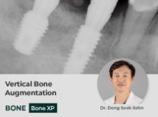
Vertical Bone Augme…
Although the shape of the defect was irregular and wide, it was visually confirmed that the partial new bone formation u…
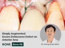
Simply Augmented Se…
New bone was created by re-using BOSS bone graft even though it was difficult to obtain initial fixation strength as the…
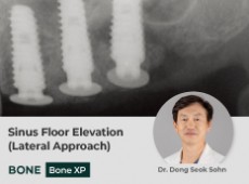
Sinus Floor Elevati…
Even though the maxillary sinus at the surgical site was inflamed, it was visually observed that the new bone formation …
Major Bone Augmenta…
The BOSS bone graft material was stably spaced despite the extensively performed vertical and horizontal bone augmentati…
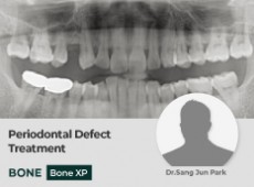
Periodontal Defect …
After 3 months of operation, the radiograph was observed similar to the tissue adjacent to the marginal bone level, and …
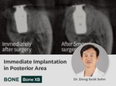
Immediate Implantat…
We observed the natural connection between the existing alveolar bone and the new bone on radiographs, and confirmed tha…
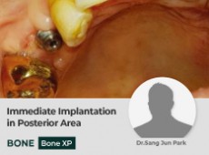
Immediate Implantat…
The implant was placed according to the root shape of the #15 tooth, and esthetic results were obtained by using POSS on…
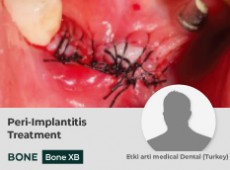
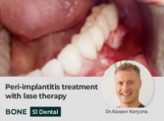
Peri-Implantitis Tr…
Form a shielding film by fixing it to the COLLA Cover screw with Poncho technic
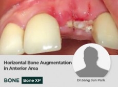
Horizontal Bone Aug…
Bone augmentation was performed in the horizontal direction, and the incision was made to minimize the scope of the oper…
COLLAGEN REGERNATION
Simplified Vertical…
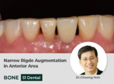
Although the range of bone loss in the defect was wide, S1 bone graft material can be applied stably to the desired area
Dough Staged Sticky…
On radiographs 3 months after surgery, it was observed that the shape of the marginal bone was harmoniously connected wi…
Immediate Implant P…
Used S1 and COLLA, the preservation of periodontal tissue was better than before extraction, and esthetically satisfacto…
Minor Bone Augmenta…
A shield was safely formed with COLLA at the surgical site, and a good prognosis was observed without inflammatory findi…
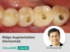
Ridge Augmentation …
A shield was safely formed with COLLA at the surgical site, and a good prognosis was observed without inflammatory findi…
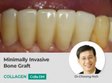
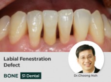
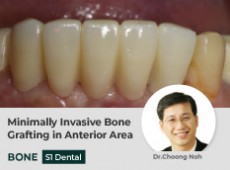
Minimally Invasive …
GBR was performed to increase the volume in the labial direction of the mandibular anterior region, and aesthetic and fu…
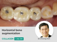
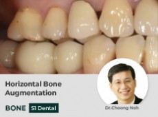
Horizontal bone aug…
Although a large amount of GBR was performed due to the buccal width of the alveolar bone, the space was maintained well…